Modern methods of medical diagnostics
CT scan
A CT scan, often referred to as a CT scan, is one of the most modern methods of medical diagnosis. It is a non-invasive test that provides detailed images of a patient's internal organs and tissues. The method is based on the analysis of X-rays and computer processing of images.
During the examination, the patient is placed on a special table that slides through a hole in the scanner. As the table moves, the scanner emits X-rays that pass through the patient's body. A detector in the scanner records the reflected rays and transmits the information to a computer. Then, using advanced algorithms, the computer generates a three-dimensional image of the examined area.
Course of ultrasound examination
Ultrasound, commonly known as ultrasound, is one of the most commonly performed diagnostic tests. This method uses high-frequency sound waves, which are harmless to the patient's body. These sounds are reflected by organs and tissues and then processed into images by specialized equipment.
During the examination, the patient lies on a table, and the doctor applies a gel-based sound-conducting agent to the area to be examined. The doctor then moves the ultrasound transducer over the skin, collecting information about the organs and tissues. The images are visible on a monitor and allow the doctor to see inside the areas being examined.
Magnetic resonance method
Magnetic resonance imaging (MRI) is a non-invasive diagnostic method that uses a strong magnetic field and radio waves to obtain highly detailed images of a patient's body. Unlike CT scans, RM does not use X-rays, making it safe for the patient.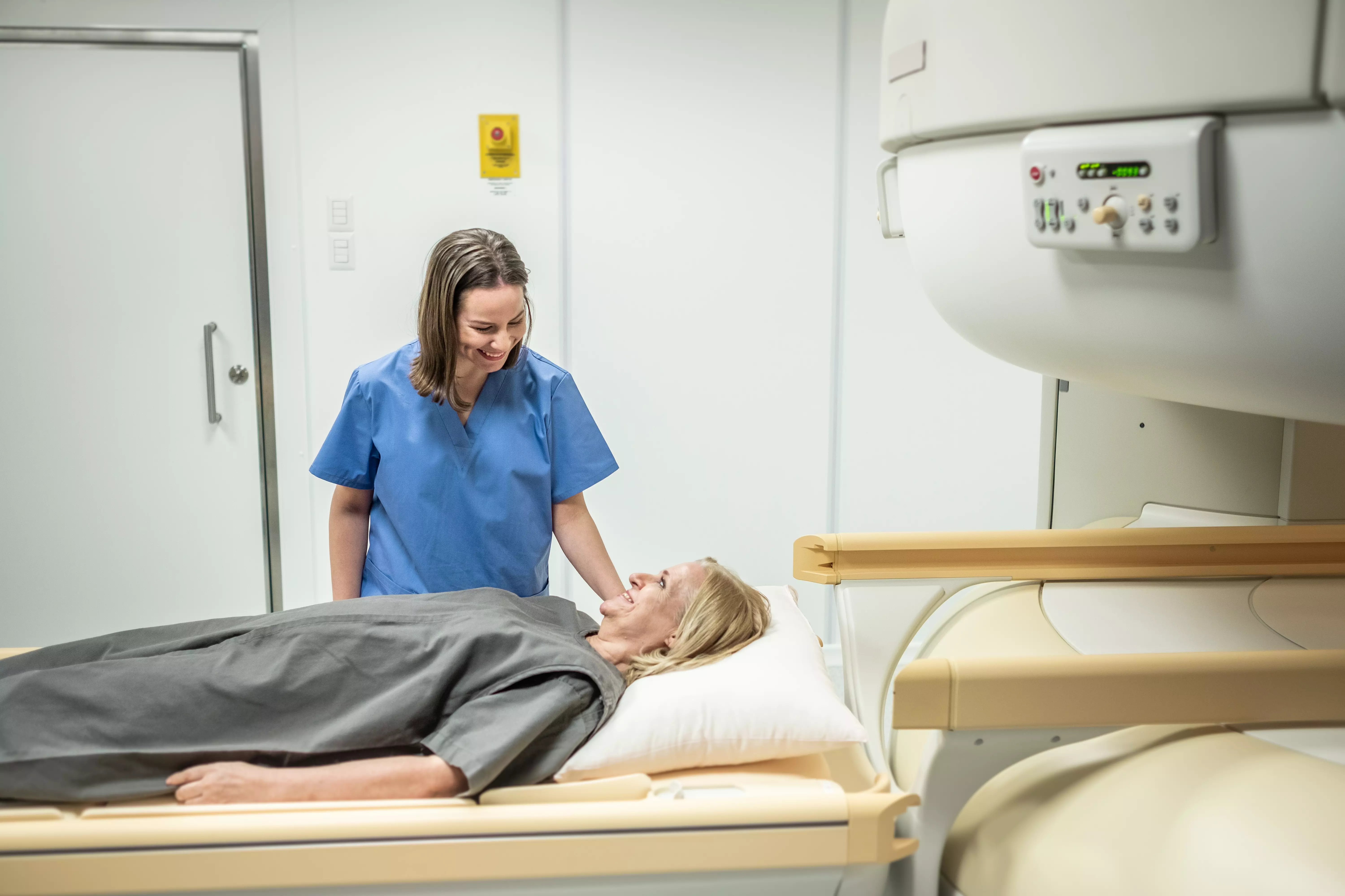
During the examination, the patient lies on a table that is moved inside the RM camera. A strong magnetic field is generated inside the camera, which stimulates the hydrogen in the patient's body. Then, different sequences of radio pulses are switched off and on, which generate electrical signals. These signals are received by special antennas and then transmitted to a computer, which generates images.
Endoscopic examination
An endoscopic examination is a diagnostic method that involves inserting a special device called an endoscope into the patient's body. An endoscope is a thin tube equipped with a camera and light that allows direct viewing of the inside of organs and various medical procedures.
During the examination, the patient is under local anesthesia or anesthesia, and the endoscope is inserted through the body's natural orifices, such as the mouth or rectum. The endoscope's cameras transmit images to a monitor, allowing the doctor to closely examine the area being examined and perform necessary procedures, such as biopsies.
Summary
Modern methods of medical diagnosis, such as computed tomography, ultrasound, magnetic resonance imaging and endoscopic examinations, allow doctors to accurately and quickly diagnose various conditions. Thanks to them, it is possible to detect diseases in their early stages, which significantly increases the chances of successful treatment. It is worth remembering that each of these methods has its own advantages and indications for use, so in case of the need for medical diagnosis, it is worth consulting a doctor who will select the appropriate method of examination.